Applications in Material Research and -testing
Spectral Analyses down to the Submicron Range
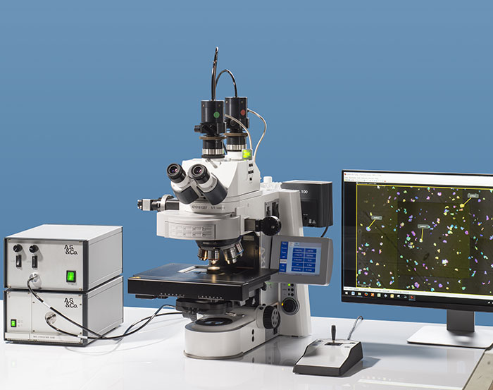
In combination with stereo- or light microscopes spectral analyses can be expanded to structures and single particles down to the submicron range. It provides information about real colors and their distribution in materials. It enables the evaluation of coating properties on glass surfaces, and via mathematical algorithms it is also possible to analyze the layer thickness of semitransparent coatings and to determine particle size and distribution in colloid substrates. This can be done directly under the microscope in a non-destructive manner. Special designed A.S. & Co. modules nd customer- specific engineering provide high sophisticated solutions that push technical possibilities to the limit.
Microscope Configurations
Light microscope spectral analyses can be done with different spectrometers in combination with various types of stereo- or light microscopes. Microscope and spectrometer are fiber connected by a collimation system designed from A.S. & Co.. Microscopes from all manufacturers can be used.
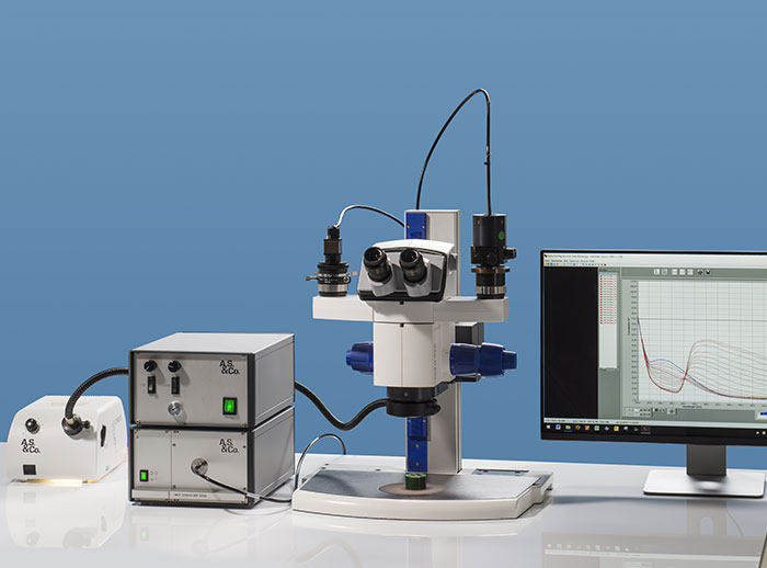
Modern stereo microscope with a double TV Cmount adapter can obtain spectra and photos from the same sample position.
© A.S. & Co. GmbH

Research light microscopes provide options for all examinations on the limits of the optical resolution, in DUV and NIR or automated spectroscopy.
© A.S. & Co. GmbH
Stereo microscopes are specifically used for samples that need a longer working distance and cases, where three dimensional view is desired. This concerns for example investigations on printed matter like books or surfaces of art objects. Stereo microscopes are easy to handle and offer a magnification range between 1 and 400x. A.S. & Co. stereo microscope configurations help to exploit the fully optical capacity of modern stereo microscopes. Manual and motorized solutions are suitable for the spectroscopy as well as flexible stands offering horizontal measurement positions. The fiber coupled layout separates the main microscope body from the sensor and avoids focus dislocation caused by too heavy device superstructures.
Conventional light microscopes using highly stabilized illumination units work in brightfield, polarization and fluorescence. They cover a lot of applications like investigations and quality control of glass, coatings and paints, determination of layer thicknesses as well as geological studies on coal and oil or maturity of sediments.
Research light microscopes equipped with manual or motorized functions are the instruments of choice for all examinations on the limits of the optical resolution or in Deep UV and NIR. Examples are the differentiation of particles in colloidal solutions, nanoparticles or structures on pigments. The forensic comparison of fibers often requires the Deep UV, because this enables differentiations between synthetic or natural fibers. NIR becomes a subject when the light penetration inside a semitransparent material shall be expanded, an application used for instance in determination of layer thicknesses and quality control of wafers.
Spectral Sensors
Compact modules implemented inside microscope controlling units are well suited for stereomicroscopic investigations and many routine tasks, for which they represent an economic solution.
Modular spectrometers systems, which combine more than 20 spectrometer modules with different types of UV/VIS/NIR sensors, cover the spectral range from 190 to 2,200 nm. They offer maximum flexibility and include DUV-NIR combinations over the complete detection ranges.
Peltier cooled CCD spectrometers provide highest possible sensitivity and cover weak fluorescence signals or darkfield results, UV and NIR 190–950 nm measurements. This high sensitivity is achieved by the large pixel size of 24 x 24 μm and the vertical binning, which exceeds the active detection area of more than 36,000 square microns per 0.8 nm resolution in comparison to a highly sensitive fluorescence CCD video camera which has a pixel size of 29 μm2.
Microscope-Spectrometer Coupling
The microscope-spectrometer coupling is the key factor for the performance. High quality results can only be obtained, when all technical and optical features are adjusted to each other.
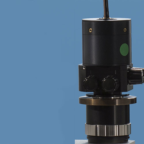
- xy-fiber alignment of the collimator
- Adjustment of pinhole size and position
- z-focusing
Microscope-spectrometer- adapter with microscope coupling, variable rectangle pinhole and collimation.
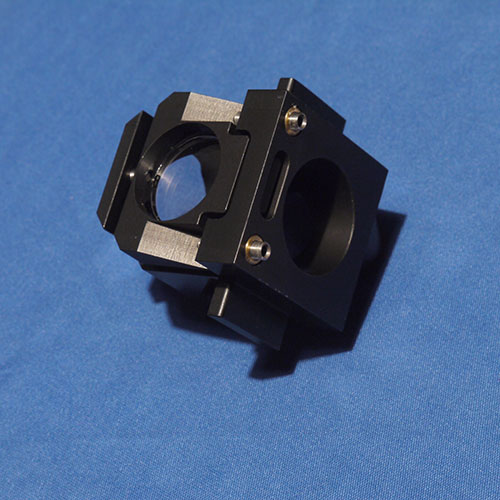
Live Overlay Module embedded in a reflector revolver module. Sample and apertures can be overlaid online through the eyepiece.
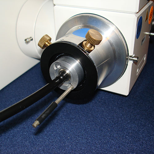
Deep UV-NIR illumination module with x/y/z fiber alignment and interchangeable optic
Special designed A.S. & Co. Microscope Collimation Adapters
- provide mechanisms for a precise x/y/z alignment of the fiber into the optical axis
- arrange the best possible optical signal transfer from the microscope to the spectrometer
- have a zoom option which projects various pinhole diameters fully into the fibers detection area
- optimize the size and position of the measurement pinhole (confocal effect)
- visualize the measuring aperture live into camera image or the eyepiece by an integrated switchable illumination
- focus the excitation light precisely to the detection point
- have an integrated shutter for automated workflow
Automated Spectroscopy with SpectraVision Mapping
Quantitative UV-NIR spectroscopy needs to meet increasing demands on high efficiency and quality standards. The workflows and technologies have to be optimized in order to minimize time and obtain sophisticated results. SpectraVision Mapping is a tool for automated spectroscopy of large samples at microscopic resolution including the full synchronization of microscope and spectrometer. It provides accelerated and high precise workflows for various applications in industry, geology, life science, forensic and art object expertise.
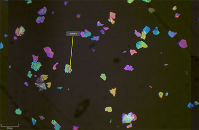
The mosaic function creates a quick overview of the complete sample. Extraction of a single tile with selected region of interest.
© A.S. & Co. GmbH
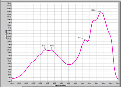
At each selected position the system records a spectrum.
© A.S. & Co. GmbH
SpectraVision Mapping allows automated spectral measurements in large area samples. Scanning and Live Stitching are the first steps in the automated process and give an overview about the samples structure. The software concatenates individual pictures taken at maximum resolution of the objective to a full screen sample overview. It is accompanied by an autofocus function, which adjusts the focal plane during the detection process. This mosaic picture can thus serve as a map to navigate through the sample and provides a basis for further analyses.
During the final positioning of the selected region of interest (ROI) the autofocus function makes a focus check for each position which is stored. This ensures that the z-detection coordinates are individually registered in submicron resolution.
Software Features
- Stitched images generated by automated scanning serve as a virtual map
- Seamless zoom-in and zoom-out without change of objective
- Selection of regions of interest (ROI) by mouse click
- Final autofocus-check to fix the ultimate measurement z-position in submicron accuracy
- Automated travel to the selected coordinates
- At each selected position the system will record the spectrum
- Automated feedback of focus and position before the measurement is started by using close loop technology for the scanning stage AND the microscope z-axis. The measurement is not executed until
- the stage has confirmed that it has reached and verified its assigned position and
- the microscope has acknowledged focus verification.
- Storage and retrieval of spectra data together with their corresponding coordinates. This enables a clear offline allocation of spectra data, corresponding coordinates and picture.
