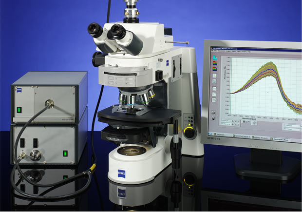
Microscope Spectrometers for Real Color Measurement
All components for UV-NIR microscope spectroscopy out of one stock

With SpectraVision A.S. & Co. provides precise spectral measurements in overall concept for complete workstations. This includes light sources for deep UV-NIR as well as cw-laser and laser diodes along with optimized microscope adaptors. Either they are mounted directly in the microscope or externally connected by a fiber collimation. Additional aperture slots or centerable luminous-field diaphragms reduce light scattering and increase the system's dynamic range.
High Flexibility of the overall Concept
The spectrometer concept developed by A.S. & Co. is open and flexible. It comprises combinable linear sensors and offers a spectral resolution down to 0.1 nm. Depending on the requirements, a workstation with a broad range of functionalities can be assembled. Combining CCD-sensors or photo diode arrays with a holographic grating inside to a monolithic miniature spectrometer
- offers a high dynamic range
- stabilizes the signal to noise ratio
- avoids time consuming on site adjustment procedures
- makes systems easy for service issues.
A.S. & Co. utilizes standard research microscopes and routine stands and modifies their optical part to extend their spectral range starting from 220 nm to 2200 nm. Limiting the application to the range between 200 nm and 2200 nm consistent, reproducible data can be easily generated over years.
For special applications a photomultiplier (PMT) can be combined with a monochromator as a microscope spectrometer in the classic style resulting in a concept comparable to the Leica MPV3 or the Zeiss MPM 800. PMT based systems offer the ability for electronic amplification obtaining a higher dynamic range in comparison to an array spectrometer. In addition, the disadvantage of undesired stray light can be suppressed more effectively by a combination of integrated blocking filters.
SpectraVision Software-Concept
The heart of our microscope spectrometer workstation is our own SpectraVision software. It comprises the following functions:
- tool-box-modules for fully automated microscope control
- adaption for requirements in daily routine
- PC-controlled measurement
- functions for laboratory needs
- functions for advanced applications
- integration of various calibration standards according to NIST or DIN 7404-5
- individual routines for microscope spectroscopy on forensic samples and petrographic studies
In addition, numerous reporting functions are available, in which either the simplest CMOS cameras for image documentation or high precision cooled CCD cameras for fluorescence microscopy may be used. They expand the measurement options by various quantitative image analysis options
Customer-specific Engineering
Customized solutions based on the combination of certified standard components with high precision special designed elements are the platform of our business philosophy.
We discuss the individual requirements for your workstation, develop a project plan and define the distribution of tasks in a system specification as a prerequisite for the offer preparation. Parallel we measure real samples to prove feasibility. Developing our own instrument control and analysis software, the A.S. & Co team is open to your ideas and inputs when the application requires software modifications.
Applications of Microscope Spectrometers
| Comparison of different structures (i.e. fibers) | Using a white standard or calibration standards in combination with defined color temperature and standardized light conditions, the spectrometer enables to display the transmission or reflectance values as a function of wavelength. Taking a spectrometer as microscope-photometer (MPM) in QC of paints and coatings, smallest inhomogeneities on pigments or in adjacent areas of a color surfaces can be differentiated; in addition it enables the comparison of different structures and colors – traditionally the forensic glass and fiber analyses makes use of this feature as comparative technology. |
|---|---|
| Rating of coal, oil and their reservoir geology | Microscopic reflectance measurements determine the market value and the suitability of fossil fuels. In addition to chemical and physical test methods they provide an essential prerequisite for the feedstock identification. The tests are standardized and described internationally through the ASTM D2798, DIN / ISO standards 7404-5:1994 and DIN 22020-5:2005. Vitrinite reflectance and spore color index comparisons also help to evaluate the genesis of petroleum source rocks and thermal maturity of the sediments in different regions. For these measurements modern light microscopes with highly stabilized illumination units can be used as brightfield, polarization and fluorescence spectrometers. |
| Differentiation of colloidal solutions and nanostructures | Microscope-spectrophotometers (MSP) also contribute significantly to the differentiation of colloidal solutions and nanostructures. Using mathematical algorithms the size of particles and their distribution can be derived from the spectra, although the individual nanoparticles are below the limit of microscope resolution. This demonstrates another advantage of high-resolution microscope spectroscopy. The sample can be characterized directly under the microscope without much effort, and it requires no additional preparation in contrast to an electron microscope. For example, a ruby red solid colored glass is usually made by adding gold colloids with a size between 2 and 100 nm to the molten glass. The size and concentration of the nanoparticles provide the ruby appearance and show spectral characteristics such as intensity peaks and pronounced half-widths that can be used for the evaluation, while the usual color impression of gold or gold-plated surfaces is missing. |
| Coating of surfaces / determination of layer thicknesses | Microscope spectrometer can also be used in the coating of surfaces and the determination of layer thicknesses, where semi-transparent materials are used. In multi-layered structure the individual coatings show different refractive indices. When light hits the boundary layer it is separated into diffraction- and reflection elements, which result in a characteristic interference pattern of the reflectance spectrum. Using appropriate simulation software mathematical fittings are capable to determine the layer thickness on a substrate in the micrometer range without damaging the structures that are being examined. This advantage of a non-destructive testing of materials is used for the study of glass surfaces or in the production of TFT displays as well as for homogeneity evaluation on plasma polymer coatings for hardening of components. |
Fundamentals of Microscopic Color Analysis with UV-VIS Spectrometers
Color analysis with a UV-VIS spectral photometer integrated in a microscope, is quite different from the RGB color model, where additive three fundamental colors combine to produce the final result. Using a UV-VIS microspectrophotometer the incident light is split into its individual spectral colors; the analysis of the absolute magnitude of the individual color shares allows measuring and quantification of the real colors in a sample. A typical example is the differentiation of blue ink of ballpoint pens, whether they consist of a homogenous blue ink or a clear carrier fluid in combination with pigments, which produce the blue color.
Real color analysis is the essential requirement for quantitative color measurements on the basis of the international CIE-LAB Norm and the comparison of standardized color values in the CIE-LAB Color charts. In contrast to simple integral color measurements, the UV-VIS microspectrophotometer allows measuring and quantifying colors in the micrometer range. From the spectral energy distribution curve the results can be processed colorimetrically according to the CIE LAB-norm and compared against reference colors charts.