Photomultiplier (PMT) for Virtrinte Reflectance Measurements
Reflectance and polarization measurements in Geology, Petrology and Economic Geology
Vitrinite is one of the main components of coal and most of the sedimentary kerogens. The measurement of vitrinite reflectance is the most important method for the determination of coal quality and usability, an indicator of thermal maturity (thermal maturity) and a tool for the characterization of Dispersed Organic Matter (DOM).
SpectraVision PMT is an efficient, easy-to-use photo-multiplier concept, which is optimized for vitrinite measurements in order to determine its content in coals or Dispersed Organic Matter. It meets the requirements of DIN / ISO standards 7404-5:1994 and DIN 22020-5:2005 and ASTM D2798 and can be attached easily to the TV port of standard microscopes. The direct PMT- microscope mounting increases the sensitivity and reproducibility of the system for fiber-coupled sensors significantly. A swivel-mounted green filter allows vitrinite and fluorescence measurements without time-consuming reconstruction or readjustment.
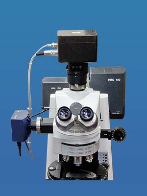
- Camera
- Direct-mounted photomultiplier
- Centering unit
- Swivel-mounted greenfilter
- Light source combination Halogen + HBO
- Rotatable polarizer
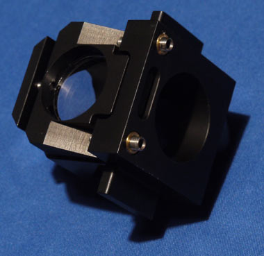
Direct-mounted photomultplier (PMT) measuring head, with centering unit for exact positioning of the PMT on a microscope’s optical axis
© A.S. & Co. GmbH
Advantages of the A.S. & Co. SpectraVision PMT
- the direct coupling to the cmount of many commercial microscopes provides a manufacturer-independent integration in existing workstations.
- the direct coupling shortens the light paths and increases the system sensitivity.
- swiveling interference filter (green) allows vitrinite and fluorescence measurements at identical sites without modification and readjustment.
- based on the sensor resolution of 16 bit associated with an electronic amplification approximately 118 000 grayscales can be displayed with the highest accuracy.
- automatic gain taking into account individual dark currents in the detection of weak light signals improves the signal / noise ratio and the level of detail.
- high degree of linearity enables the recording of areas with large differences in brightness.
- Spectral range between 195nm and 800nm
- automatically controlled shutter protects the sensor from excessive irradiation
- For measuring very small structures an additional external pinhole with integrated white light illumination can be fixed in the beam path. It allows optimal positioning of the sample while avoiding any misalignments, because there are no parts to be moved in the beam path.
Field Stop Aperture module with integrated LED white light illumination and fiber collimation externally fixed at the microscope
© A.S. & Co. GmbH
System Configuration
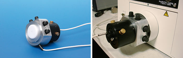
Photomultiplier and preamplifier are assembled to form a unit that can be mounted either directly on the microscope or integrated in a 14 inch housing for use externally via an optical fiber.
The system is protected from excessive irradiation by an electronic shutter. A pilot lighting integrated as standard allows easy centering on the optical axis of the microscope.
The power supplies required to operate the PMT are provided by the microscope controller, which concurrently is able to perform numerous functions in order to integrate manual microscopes in automated measurement procedures. For example, an attenuation filter can be automatically swiveled into the beam path for selecting the sample position. By the use of attenuation filters light intensity is reduced without reducing the light source voltage. Thus, a constant, firmly defined measuring and monitoring illumination is ensured.
Also further use of existing systems (e.g. Carl Zeiss MPM 800 or UMSP 80 LEITZ MPV or Craic Coal Pro) can be achieved with new software and hardware.
Microscope controller with control functions for a direct mounted photomultiplier unit
© A.S. & Co. GmbH
Specials of the Microscope -PMT Coupling
A. S. & Co. offers the opportunity to mount the PMT directly on the TV output of many commercial microscopes. The result is an increased light yield. Reflectance values in the range of ≤ 0.2% become significantly displayed and the measurement errors remain below 1%.
A swivel-mounted green filter can be optionally integrated above the phototube. Thus, the beam path to the eyepiece is kept open. As a result the sample can be observed in the white light and be measured with IF green filter without any additional step. If fluorescence measurements will be performed, the filter is swiveled-out without centering the beam path again. This makes the handling for simultaneous vitrinite and fluorescence measurement extremely convenient.
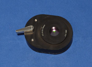
Swivel-mounted green filter above the phototube
© A.S. & Co. GmbH
Aperture systems with variable iris or interchangeable fixed apertures between 5 microns and 2 mm define the measuring area in the microscope image. Thus standardized conditions can be established quickly and easily. For optimum alignment with the optical axis of the microscope, a centering unit was incorporated in the cmount. The measuring area can be reflected into the eyepiece via a rear aperture and adjusted with the help of a crosshair, so that its location is known during the centering of the sample.
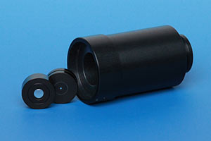
The PMT-Adapter contains interchangeable apertures with fixed diameters to establish standardized conditions in a quick and easy way.
© A.S. & Co. GmbH
Modified spectro phototube: The standard phototube was optimized to a higher light throughput. Since all optimizations potentiate, the improved light throughput at the tube has a positive impact on the overall performance and reproducibility.
Together with the A. S. & Co. product "Live overlay module" the illumination pinholes and measuring pinholes are reflected direct to the eyepiece. The viewer observes the sample image as well as the aperture, its position, size and sharpness simultaneously live. It requires no extra offline camera image on the monitor, which needs to be re-calibrated each time you change the magnification.
Live Overlay element embedded in Hartmut Althoff reflector revolver module
© A.S. & Co. GmbH
SpectraVision System Control
A PC-controlled illumination management ensures optimal modulation of the dynamic by means of neutral density filters without the need to change the lamp voltage. Maintaining absolutely constant lighting conditions are basic for accurate photometric results and achieve an optimal signal/noise ratio.
External shutter and filter changer modules allow the integration of older, purely mechanical designed microscopes into an automated measurement procedure. Likewise SpectraVision can replace the older software systems such as the Zeiss UMSP or MPM or the Leica MPV.
Motorized microscopes facilitate the workflow and can be easily integrated.
As an option motorized stages can be integrated into the process for individual mapping functions.
In systems with automatic dynamic check the modulation is made to the best use of the linear part of the sensor bandwidth.
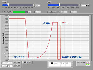
PC controlled automatic recording of gain and offset for optimal sensor dynamic
© A.S. & Co. GmbH
Additional Hardware Options
- external double monochromator for spectroscopy with maximum dynamic
- CCD spectrometer as a fast option for the recording of spectra
- Transmission equipment for displaying spectra of palynofacies
- digital camera systems for documentation and image analysis
SpectraVision PMT Software
Features for controlling and optimizing the photomultiplier measurement
- automatic microscope control
- online display of dark current
- online automatic gain and offset
- automatic gain of weak signals
- automatic adjustment of the dark current correction for gain change
- setting of different evaluation modes
- calibration with up to 15 reflectance standards
- calibration using the table values or against mathematically determined reference values and curves
Results Discussion
- display of histograms with intensity, reflection and absorption values corresponding to the applicable DIN / ISO / ASTM standards
- simultaneous online calculation of mean values and standard deviations
- standardized plots of the histograms and spectra as well as their bitmap conversion
- table view of the numerical results
- When using additional spectrometers:
Routines for normalization of spectra, for the calculation of mean values of spectra, their variances and tolerance ranges, as well as automatic peak finding routines for quick display of calibration peaks of gas discharge lamps or for the determination of the maxima - Export to Excel and ASCII
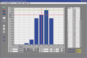
The SpectraVision window provides all necessary information about the histogram, tables of numerical results, means and standard deviations with one click.
© A.S. & Co. GmbH