Microscopic Analyses in Forensic Science
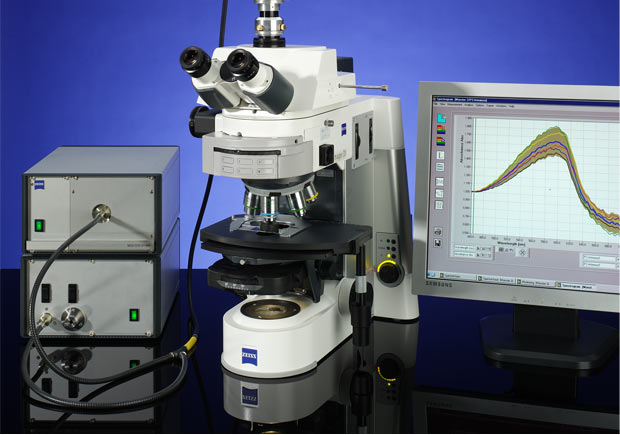
Quick and easy Microscopy for daily Routine Work
Light microscopic evaluations with AS & Co. SpectraVision are fast and economic because they allow:
- non-destructive examination of evidence
- the comparison between an unknown sample and known references
- the acceleration of routine examinations by measuring the entire spectral range within a few seconds, and
- to draw conclusions about an added value of deeper analyses
The SpectraVision concept of A. S. & Co. combines motorized microscopes with:
- various illumination systems
- photometric analysis
- state of the art sensor technology
as well as hardware and software modules that can be combined individually on request.
Suitable for Deep-UV NIR from 250 nm to 2200 nm
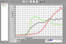
Distinction of silk, wool and polyester by comparing Deep UV result between 250 and 450 nm
© A.S. & Co. GmbH
When trying to differentiate between natural fibers and similar-colored polyester fibers in the classical spectral range of visible light there is hardly any difference noticeable; however under UV light below 310 nm the differences are clear and significant.
For an extended measurement range from 200 nm to 2200 nm with standard research microscopes, comprehensive changes on its optical properties are necessary. A.S. & Co. microscope spectrometers are modified to match these customer requirements:
- the Koehler illumination is adapted
- we utilize deep UV transmission that covers the required range down to 200 nm
- we combine various sensors optimized for a micro-spectro-photometer.
Adjustment with maximum Precision by special Illumination and Pinhole Design
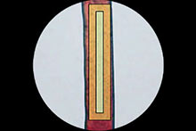
Arrangement of two rectangular apertures for the precise measurement of a longitudinally-oriented red fiber. They only cover the sample area and reduce stray light in the best possible way.
© A.S. & Co. GmbH
A longitudinal shaped fiber in a transmission beam path represents a classic problem in quantitative microscopy. The overall white light illumination of the observation field outshines the color information of the longitudinally-oriented sample. This causes a significant reduction in contrast. SpectraVision microscope adapters from A.S. & Co. use various illumination options and aperture setups for their systems avoiding this problem. The sample area is illuminated exclusively, thus minimizing over-illumination. In consequence the full dynamic range can be used to get the best spectrum.
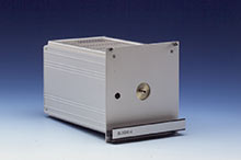
Perfect Illumination with fiber-coupled Light Sources
The centering of illumination is typically very time-consuming and requires experience with the microscope to meet the demands of precise measurement with the lowest possible error- tolerance. SpectraVision from A. S. & Co. uses highly stabilized, fiber-coupled illumination, thus meeting the requirement for standardized conditions. The light source is factory prealigned and can be exchanged or replaced without any further adjustment.
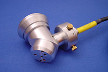
Lamp cassette with pre-centered illumination unit that can be directly connected to the beam-path via SMA Fibercoupling.
© A.S. & Co. GmbH
Optical fiber illuminator with expansion and centerable reflection mirror for DUV fullfield illumination with imaging systems.
© A.S. & Co. GmbH
Broadband Spectrum by a Combination of Light Sources
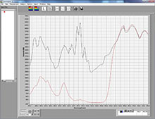
Standard spectrum of a commercially available Deep UV-NIR lamp combination (black) connected to a microscope. The typical integration time in Microscope Spectrometer from A.S. & Co. is in the range of 250 ms when Deep UV Koehler illumination is used. A result of this reference is the spectral response of a polyester fiber displayed in red. In this curve the slot between 500 and 580 nm shows impressively that any form of straylight is suppressed by the optical layout of A.S. & Co. microspectrometers
© A.S. & Co. GmbH
The combination of illuminations with different characteristics like a deuterium module with a halogen light source is one suitable configuration for microscope spectroscopy. It results in a broad spectral range of from 200 nm to 2200 nm, and ensures a stable measurement setup for years.
More benefits are:
- the increase of the Deep-UV intensity share in comparison to classic XBO illumination
- the compensation of strongly-pronounced peak maxima in gas discharge lamps by wavelength optimized conversion filters and mixing with the halogen contribution
- an increase of energy to a relatively uniform excitation spectrum, exploiting the full dynamic range of the sensor more efficiently
- the reduction of the total radiation intensity by the use of more sensitive Peltier cooled detection Arrays
- the significant increase in the lamps optical signal/noise stability
The beam paths will be aligned and verified by the use of pilot lights in order to protect microscope and samples from short wave DUV radiation damage. Integrated PC controlled shutters are standard to limit DUV exposure to the minimum time required for the measurement only.
SpectraVision
- integrates the synchronization of the pilot light and measurement illumination
- enhances the long-term stability and lifetime of the workstation
- reduces damage to the samples
- protects the user from DUV radiation
Free Choice of Microscope modules for Research and Routine Work
SpectraVision from A.S. & Co. uses the TV port on the microscope, which is standardized on each device. Depending from the microscope configuration all contrasting methods may be available. The microscope illumination allows the positioning of the sample, while the photometer-lamp combination ensures constant and reproducible measurement conditions in:
- Brightfield transmission
- Darkfield transmission
- Polarization
- Incident light reflectance
- Incident light darkfield
- Incident light polarization
- Fluorescence
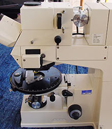
Upgrade of an older microscope photometer Carl Zeiss MPM 800 into a dual operation unit with classical monochromator-PMT measurement as well as modern illumination accompanied by cooled CCD Array Technology. This allows the direct comparison between old and new results on the same sample position.
© A.S. & Co. GmbH
The development of mobile solutions or the integration of a spectrometer into stereo microscopes is feasible. Furthermore established reference spectrometer like LEITZ MPV 3 or Zeiss UMSP 80 can be upgraded also with new sensor technology, modern computer and up to date software. The upgrade also integrates established high precision aperture mechanics, combines them with the characteristics of UV-capable modules and the advantages of modern electronics.
Quantitative Fluorescence Measurements
Fluorescence signal from a banknote, illuminated with 365 nm.
© A.S. & Co. GmbH
In the past quantitative fluorescence measurements were disadvantaged by the unavailability of calibration standards and by time consuming measurement procedures. The observed samples were negative affected by the long and intensive radiation leading to poor reproducibility achieved.
One main focus of SpectraVision is the objective to achieve highly reproducible fluorescence results. In cooperation with scientists we established a calibration method, which compares the fluorescence intensity versus an intensity spectrum of a Tungsten lamp. Combined with the knowledge of various instrument parameters we create valid fluorescence quantification.
SpectraVision from A.S. & Co. optimized for microscopic fluorescence spectroscopy uses
- cooled CCD-spectrometers with short exposure times for the full spectral range
- a dynamic range up to 16-bit
- optical microscopes with apochromatic, color-corrected beam paths
- additional accessories and apertures for a scatter-free beam guidance
- the newest interference filter combinations
- monochromators for the flexible selection of excitation wavelengths
- calibrated light sources with defined peak maxima as a reference
Optionally, photomultipliers and avalanche diodes can be added as sensors for high-sensitive luminescence measurements or as amplification systems to enhance weak signals.
Polarization Spectroscopy to analyze Birefringence
Polarization microscope spectroscopy, which shows birefringence on fibers, crystals and mineral structures or characteristics of biological materials, can also be utilized for the differentiation of trace compounds. In this case the evaluation of color- and brightness portions as a function of the rotation angle is the basis for the differentiation.
SpectraVision from A.S. & Co. uses the information of polarization microscopy by
- recording of minimum and maximum reflectance as a function of the rotation angle
- measuring spectral changes with an online monitoring function
- recording of spectra and their evaluation against reference curves, which are taken from literature sources or were measured against calibration standards
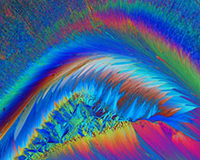
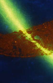
Vitamin C excited by crossed polarizers
Foto: Frank Brückner, LKA Eberswalde
Combining different Methods
UV-NIR spectroscopy has been one of the standard methods in forensic investigations for decades. The main application is the comparisons of fibers, paint and coatings. As nowadays comparisons of groups of substances are often made at the molecular level using methods of FTIR or Raman spectroscopy, UV-VIS technology finds new applications in
- the examination of microstructures in pigment surfaces,
- the non-destructive analysis of layer thickness
- the measurement of surface reflectance
- the rapid testing of the sizes of nanoparticles or their distribution in colloidal solutions.
UV-VIS fluorescence spectroscopy combined with image processing provides information on the composition of printing colors and inks; color measurements and particle analysis support the identification of carrier solutions of an ink or they distinguish suspensions through structural characteristics and particle measurement.
A 3D documentation of color penetration with information on the changes of the colors due to thermosublimation enables conclusions about the printing technique. Furthermore the age of questioned documents can be examined through the diffusion of pigments into the surface of the paper or through the examination of chemical changes in the color.
NIR microscope spectrometers are used for the rapid classification of NIR markers in document papers. They can serve as cost-effective sensors for routine testing.
All the described spectroscopic methods benefit from the advantages of fast, non-destructive microscopic analysis. The flexible concept of SpectraVision Hard- and Software from A.S. & Co. allows the free combination of UV-NIR microscope spectrometer modules with FTIR, Raman or confocal systems integrated in research grade microscopes with highest resolution lenses.